
Diagnostic Tests
Electrocardiogram/EKG
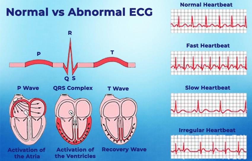
An electrocardiogram, also known as ECG or EKG, is a basic, harmless test that detects and records your heart’s electrical activity. An EKG shows both the rhythm of your heartbeats, whether they are normal or irregular, and the strength and timing of the electrical impulses passing through each part of your heart.
Your doctor may order an EKG to assess:
- Your heart rhythm, determine if it’s normal or if you have an arrhythmia
- Poor blood flow
- A heart attack
- Abnormalities of your heart, such as heart chamber enlargement and abnormal electrical conduction
- Heart damage or heart failure
- Heart condition for upcoming surgery
- Effects of new medication for heart disease
There are little to no risks for an EKG test. There is no radiation or electricity put into your skin, but you may experience some skin irritation after removing the electrodes.
You can eat and drink like you normally would before your EKG test. You will need to be mindful of applying oily or greasy skin creams or lotions before your test. This could interfere with the electrodes' ability to stick to your skin. Also, take note to avoid full-length hosiery as electrodes will be placed on your legs. To ensure a smooth process for your EKG test, it’s best to wear a shirt that you can remove easily to place electrodes on your chest.
This test can be performed in different places such as a doctor’s office, an outpatient facility, at a hospital before surgery, or as a part of stress testing. You will lie flat on a table or patient bed. A nurse or technician will adhere electrodes to your chest, arms and legs. As a precaution, any body hair preventing the electrodes from sticking may need to be shaved. These electrodes, connected to the machine by wires, then record your heart’s electrical activity. Once the test is completed, the electrodes are removed.
References
Cardiac Event Monitor
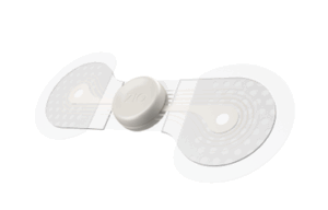
A cardiac event monitor is a wearable device that records your heart rhythm and rate continuously for 24 to 48 hours or longer. Compared to an EKG, a cardiac event monitor can provide more information on your heart's electrical activity. The purpose of an event monitor is to record your heartrate and rhythm during an "event"/symptom. Abnormal heart rhythms and symptoms can come and go therefore a cardiac event recorder can best capture your symptoms. Whether you have a fast, slow, or irregular heartbeat, your doctor may ask you to wear this device. A cardiac event monitor can also help find the cause of arrhythmias, heart palpitations, and unexplained dizziness.
There are no risks with wearing a cardiac event recorder, however, the adhesive from the electrode patches can cause some irritation. Applying a cardiac event recorder is simple, you may either apply it yourself or have a technician do it. As a precaution, your skin must be free of oils and creams. If you are to apply it yourself, you will receive instructions along with the device. Other cardiac event recorders can be held or worn on your wrist and do not require electrodes attached to your chest.
There are different types of events recorders such as an implanted and external loop memory monitor, symptom event monitor, and patch recorders. All devices can send your EKG readers to a transmission or receiving center. The EKG data will be analyzed and reviewed by a Certified Cardiographic Technician who will then provide a report back to your doctor.
Echocardiogram
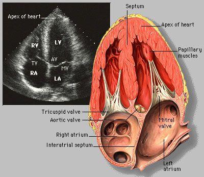
An echocardiogram, also referred to as an echo, is a pain-free test that uses sound waves to create moving pictures of your heart. The pictures depict the size and shape of your heart, and how well your heart is pumping blood. An echo can also detect blood clots inside your heart, fluid buildup in the pericardium (the sac around the heart), tumors and issues with the aorta. The aorta is the main artery that carries oxygen-rich blood from the heart to the body.
Your doctor may order an echo for several reasons. You may simply be taking this test if you are preparing for surgery or a procedure. If you are experiencing symptoms, your doctor may order this test to learn more about your heart. One reason may be to find the cause of abnormal heart sounds like heart murmurs that are due to damaged heart valves. Another reason is to see how well your heart responds to certain treatments or to check on a condition you’ve already been diagnosed with.
There are three different types of echocardiograms:
- Transthoracic echocardiogram (TTE)
- Transesophageal echocardiogram (TEE)
- Fetal echocardiogram
- Stress echocardiogram
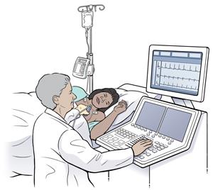
The standard echocardiogram ordered here in our medical office is the Transthoracic echocardiogram (TTE). A TTE is a noninvasive way to look at blood flow through the heart and heart valves. This test creates images of the heart from outside the body. Contrast may be given by IV to help the heart’s structures show up better in images.
A standard echocardiogram poses no known risk to the body and there is no X-ray exposure. You may feel some discomfort when the transducer pushes against your chest. This firmness is necessary to create the best images of your heart. On the contrary, there is risk for some people who may get a reaction from the contrast dye. Most common reactions would be backaches, headaches, or rashes and these typically happen immediately after administering the dye. Severe allergic reactions are rare.
There is minimal preparation for a TTE, you can eat or drink as you normally do before your test. Ensure your healthcare team knows about all the medications and supplements you take.
This test can be performed in a medical center or a hospital. You are only required to undress from the waist and up. The technician will put gel on the transducer, press it firmly against the skin and move it across your chest. The transducer sends sound waves through the chest to the heart and records. The computer will transfer these recordings into moving images.
References
Stress Echocardiogram
A stress echocardiogram is part of a stress test. This test is used to see how well your heart handles work. As your body works hard during the test, it requires more oxygen, resulting in the heart pumping more blood. This test will show if the blood supply is reduced in the arteries that supply the heart.
Your doctor may order this test if you have symptoms of heart disease, specifically if they worsen with activity. Some symptoms include:
- Shortness of breath
- Dizziness
- Rapid or irregular heartbeat
- Chest pains
A stress echo is safe and has few risks. Risks you may experience are due to your underlying heart condition. When you stress your heart, you can experience an abnormal heart rate or chest pain and pressure. You will be monitored closely for signs of distress and the test will immediately stop if necessary.
A stress echo is safe and has few risks. Risks you may experience are due to your underlying heart condition. When you stress your heart, you can experience an abnormal heart rate or chest pain and pressure. You will be monitored closely for signs of distress and the test will immediately stop if necessary.
You will receive instructions from your doctor on preparation for your test. It is important to avoid caffeine 24 hours before the exam. No smoking or using tobacco day of the test as well. Do not eat or drink within the hours before your test. Make sure to dress comfortably as you will be exercising.
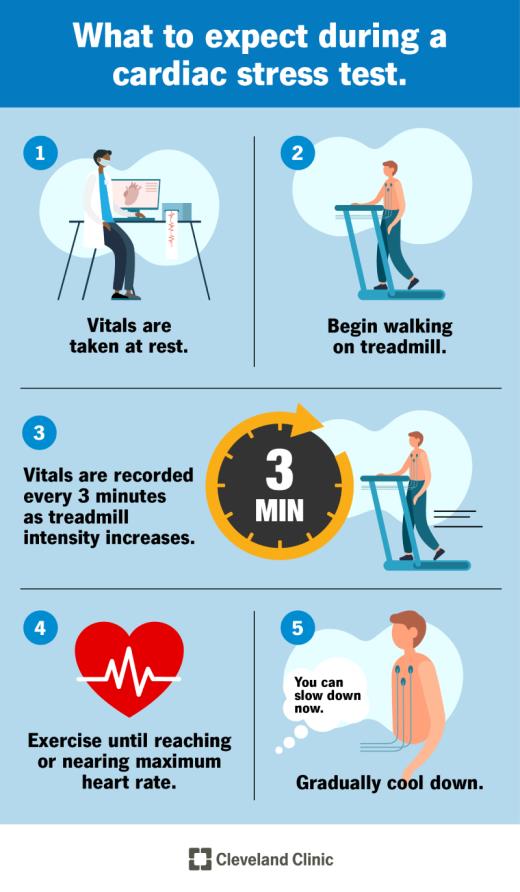 There are two types of stress tests: a treadmill/exercise stress test and a dobutamine stress test. For an exercise stress test, you will be asked to exercise on a treadmill. This test can be performed in a doctor’s office or a hospital. Before you begin your exercise, a technician will place electrodes on your chest, attach a blood pressure cuff to your arm, and a pulse monitor to your finger or another part of your body. The technician will then measure your heart activity and blood pressure before you start the test. When the test begins, you will walk at a slow pace and then gradually increase the treadmill speed until your heart rate is at the target rate for your age. Your heart’s electrical activity, heart rate and blood pressure may also be measured during your exercise. The total duration of the exercise is about 10-15 minutes; however, the test will stop if you show any sign of a heart problem or if you are too tired to continue the test.
Your technician will also use echocardiography to capture pictures of your heart during or after the stress test. Once the test is complete, your technician will measure your heart activity and blood pressure again to ensure that both measurements are back within normal range. It’s expected that you can return to your normal activities after performing this test.
If you are unable to exercise, your doctor will likely order a dobutamine stress echocardiogram. This test can be done safely in an outpatient clinic or a hospital. Your doctor may order this test to assess:
- The heart’s function and structure
- The degree of known heart valve disease
- Limits for safe exercise or recovery from a cardiac event
- Cardiac status before heart surgery
Possible risks related to the dobutamine stress echo include:
- Severely high blood pressure
- Irregular heartbeats
- Chest pain
- Dizziness
- Nausea
- Heart attack (uncommon)
When preparing for your dobutamine stress echo, you will be asked to sign a consent form that gives permission to do the test. Inform the doctor if you are allergic or sensitive to any medicines or latex. Fasting may be required before the test. You will receive instructions should you need to withhold food and liquids.
Similarly, it is important to tell your doctor all medications, vitamins, herbs and supplements you are taking as you may be instructed to hold certain medications before the test. It is important to note that dobutamine will make your heart beat fast and mimic the effects of exercise, so smoking and ingesting caffeine before this test may interfere with the dobutamine. If you are pregnant, you could be pregnant, or have a pacemaker inform your doctor.
When you come in for your test, you will be asked to remove any jewelry or objects that may interfere with the test. If you use glasses, dentures, or hearing aids you can keep those items on during the test. You will be asked to remove clothing from the waist up and wear a gown. Then you will be asked to empty your bladder before the test. An intravenous (IV) line will be put in your hand or arm before the test for injection of the dobutamine and IV fluids, if needed.
 You will lie on your left side on either a table or bed but may be asked to change position during the test. You will be connected to an ECG monitor to record your heart’s electrical activity. Your vital signs will also be monitored during the test. The ECG tracing will be compared to the images displayed on the echocardiogram monitor. The technologist will place a warmed gel on your chest and then place the transducer on the gel to capture an image of your heart. The dobutamine infusion will begin at a rate determined by your weight. The infusion will increase every few minutes until you have reached your target heart rate or until the maximum dose of dobutamine has been reached.
When the dobutamine starts and after each increase, your blood pressure will be checked, an ECG tracing and echo will be recorded. The technologist will get images of all areas and structures of your heart. When you have reached your target heart rate or the maximum amount of dobutamine, the medicine will be stopped. Your vital signs will continue to be monitored for 10 to 15 minutes until they have returned to the baseline state. The final echo images will be taken, and the test is complete. The technologist will wipe the gel, remove ECG pads, and take out the IV line. It is important that at this stage if you feel any chest pain, trouble breathing, sweating or heart palpitations please inform the technologist.
References
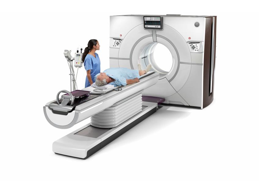
Cardiac CT
A cardiac computed tomography scan (CT scan) is a three-dimensional image of your heart, vessels and surrounding structures. With a CT scan, your doctor can see coronary arteries, heart chambers, pulmonary veins, thoracic aorta, and your pericardium.
This procedure is done in a hospital or an imaging center. Your doctor may need you to do a cardiac CT scan to assess:
- The cause of chest pain and shortness of breath
- Your heart arteries for calcium buildup, narrowing or blockages
- Overall, your heart valves
- Any problems with your aorta, aneurysms and dissection included
- The sac around the heart, for fluid or calcification
- And plan for an arrhythmia ablation procedure
A CT scan has minimal risk, some patients may experience reactions to the dye. This includes itching, nausea, sneezing, or a rash at the injection site. Symptoms will go away without treatment, however antihistamines, for example, can help. It is rare for serious allergic reactions to occur, but few may experience an anaphylactic reaction resulting in difficulty breathing. If you are diabetic or have kidney disease you may need to drink more fluids post-scan. This will help flush out the contrast agent, iodine, out of your body. For women breastfeeding, it is best to pump and prepare milk for your baby before you receive contrast as it can get into your breast milk.
Since CT scanners use X-ray, there are small risks of exposure to radiation. The amount of exposure to radiation should be kept to a minimum. For pregnant women, radiation can be harmful to a fetus, so this scan is not recommended.
There are detailed instructions you’ll need to follow before and on the day of your CT scan, your healthcare team will send those to you. Day of your scan do not eat for four to six hours before your appointment time. You may drink water but avoid caffeinated beverages such as coffee, tea, energy drinks or sodas.
When you come in for your CT scan you will change into a hospital gown. If necessary, you will be asked to take medicine that slows down your heart rate. If you are ordered a test with contrast, a nurse will insert an IV line in your arm to administer the dye. You will lie flat on the CT bed. The technician will place three electrodes on your chest to monitor your heart’s electrical activity during the scan. The entire scan and preparation usually last between 30 to 60 minutes.
References
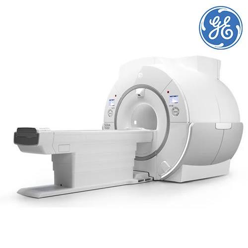
Cardiac MRI
Cardiac magnetic resonance imaging (MRI) uses radio waves and magnets to create images of your heart. Unlike other cardiac imaging, an MRI does not use radiation instead
the radio waves and magnets detect molecular changes in your body. It can show all parts of your heart, how well they function, and the blood flow. MRI scans are also much more accurate and provide detailed images of your heart from different angles.
Your doctor may order a cardiac MRI to:
- Determine the cause of your heart symptoms (i.e. chest pain, shortness of breath, or fainting)
- Navigate location needed to treat during an ablation
- Prepare for a procedure or treatment
- Assess effect of treatment on your heart/success of recent surgery
- Evaluate heart issues (muscle damage, infection, protein buildup, iron deposits, inflammation)
- Check for damage after a heart attack or lack of blood flow from artery blockages
- Diagnose heart failure, congenital heart disease, heart masses, valve disease, irritation of the pericardium
 A cardiac MRI is harmless but if you are pregnant or have metal in your body, this scan may not be recommended. The MRI machine can also interfere with medical devices while you are inside. Make sure to tell your doctor what medical device you have, they can double check if your device is safe and compatible with the MRI machine. If you have other metals in your body, the MRI machine can make the metal move or heat up. If you receive contrast, you may experience a metal-like taste in your mouth for some time. You may also experience bruising or irritation at the injection site of the IV. On rare occasions, patients experience headaches, nausea, or an allergic reaction to the contrast.
To prepare for a cardiac MRI, you will want to notify your doctor of any allergies, health problems, and past surgeries. If you have low kidney function, your doctor may not give you contrast for your cardiac MRI as it may be difficult for you to clear the contrast dye from your body. If you are claustrophobic, you can ask your doctor a few days in advance for a sedative. Ensure you will have someone to drive you to and from your cardiac MRI appointment, if you are going to be taking this sedative.
References
Myocardial Perfusion Imaging Test
Myocardial perfusion imaging (MPI) is a non-invasive imaging test that depicts the blood flow through the heart muscle and how well the heart muscle is pumping. Areas of the heart that are not getting enough blood flow can be seen on this test.
There are two kinds of MPI: positron emission tomography (PET) and single photon emission computed tomography (SPECT). The difference between the two is the radioactive tracer used during the procedure. A PET scan of the heart uses nuclear medicine. Radioactive tracers produce pictures of your heart and can show healthy and damaged heart muscle. Doctors use PET scans to diagnose coronary artery disease (CAD) and damage due to a heart attack. A radioactive tracer is injected, which created the image of your heart by tracking how your body reacts to the tracer.
A SPECT scan of the heart includes a small amount of radioactive tracer being injected into the bloodstream. The tracer produces energy inside your body. A camera picks up signals from the tracer, and a computer converts them into pictures of blood flow through the heart.
Most patients who experience chest discomfort will undergo this test to determine if the pain, called angina, is caused by the lack of blood flow to the heart muscle. This lack of blood flow can be caused by narrowed or blocked heart (coronary) arteries. This test can also show if you previously had a heart attack.
Your doctor may also order this test for you if:
- You need to determine how well your heart can handle physical activity
- Have heart damage from a heart attack
- You should have a coronary angiogram
- You would benefit from a coronary stent or bypass surgery
You may be exposed to a low dose of radiation, but generally MPI tests are safe. If you’re pregnant or think you might be pregnant, or if you’re a nursing mother, tell your health care professional before you have this test. It could harm your baby.
In preparation for this test, you need to tell your healthcare provider about any medications you take, including over-the-counter medications, herbs and vitamins. Do not consume caffeine-containing beverages or chocolate 24 hours before your test if instructed not to. You may also be instructed to avoid eating or drinking after midnight before your test.
This test can be performed in a hospital or clinic. A trained technician will place small pads (electrodes) on your chest, arms and legs. The pads have wires that hook up to a machine to record your EKG. The EKG keeps track of your heartbeat during your test and is used to tell the camera when to take a picture. You will wear a cuff around your arm to track your blood pressure. Your technician will put an intravenous line (IV) in your arm, and you will exercise on either a treadmill or a stationary bicycle. If you are unable exercise, you will do a chemical stress test. Your IV will be connected to a bag that has a medicine to widen the arteries in your heart or make it go faster, similar to when you exercise. When you have reached your peak activity level, you’ll stop and receive a small amount of radioactive material (tracer) through the IV. You will lie still on a table, with your arms above your head, for 10-30 minutes while the gamma camera takes pictures of your heart. Several scans are taken to provide pictures of thin slices of your entire heart from all angles. During the resting part of the test, you’ll receive more tracer, and another set of pictures will be taken. This set of images will be compared to the images taken after exercise or stress. Some forms of the test don’t use stress or exercise but take sets of resting images with a tracer.
After this test, you may return to your normal activities. Drink plenty of water to flush the radioactive material from your body.
References